3D NLS-graphy and computer tomography in diagnostics of benign tumors of greater salivary glands
143 patients with greater salivary gland tumors were examined, all of them underwent 3D NLS-graphy, 64 patients underwent non-contrast computed tomography (CT). 78 pleomorphic adenomas were registered in total. In 56 (71.8%) cases, adenomas were located in parotid salivary glands, in 22 (28.2%) cases adenomas were found in submandibular glands. In 46 (82.2%) cases, 3D NLS-graphy presented pleomorphic adenomas of parotid salivary gland as oval or round hyperchromogenic formations with distinct contours and heterogeneous structure. Vascular bed affection was registered in 74 (94.9%) of pleomorphic adenomas cases according to 3D NLS-angiography. Computed tomography presented pleomorphic tumor of parotid salivary glands as high-density single formation with round shapes, distinct lines and even contours in 44 (93.6%) cases.
3D NLS-graphy demonstrated 100% of sensitivity, 96.3% of specificity and 97.7% of accuracy in detection of pleomorphic adenomas of large salivary glands; CT demonstrated 97.6, 96.4, 97.6% respectively in doing so. 3D NLS-graphy is the main imaging method.
Introduction part
Frequency of large space-occupying lesions of salivary glands in China reaches up to 7% from all diseases of maxillofacilal area. Pleomorphic adenomas are the most common tumors, making from 50% to 65.7% (according to various researches) of all space-occupying lesions of large salivary glands. Diagnostics of such adenomas presents certain difficulties due to uniformity of clinical and instrumental diagnostic signs; as a result, diagnostics error rate varies from 7 to 46%.
The goal of given study is improvement of diagnostics method of large salivary glands pleomorphic adenomas using 3D NLS-graphy and non-contrast computed tomography (CT).
Material and methods
143 patients with tumors of greater salivary glands (age group from 15 to 78 years) were examined to determine the capabilities of given methods. There were 103 women and 40 men patients. All patients underwent 3D NLS-graphy and 64 patients underwent CT of salivary glands procedure.
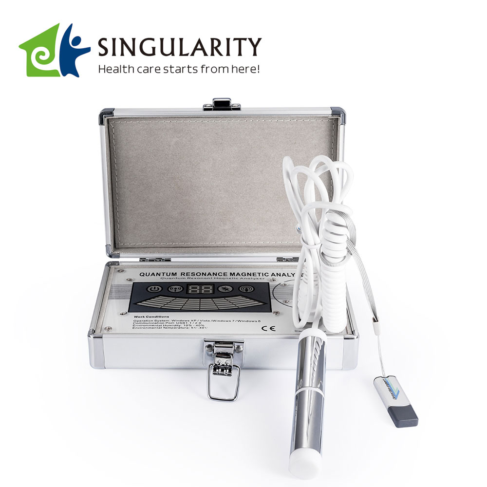
3D NLS-graphy of salivary glands was carried out according to standard procedures. 3D NLS-graphy was performed using «Metatron»-4025 (IPP, Russia) system with «Golden Dragon» software and the possibility of three-dimensional (3D) visualization of salivary glands.
Intratumoral vascularization was estimated according to 6-point Fleindler’s color scale (from 1 to 6 points) when using 3D NLS-graphy in space-occupying lesions of salivary glands patients. Spectral-entropy analysis was simultaneously performed which allowed estimating the nature of salivary gland neoplasms.
Non-contrast CT of large salivary glands was performed using Ligth Speed (General Electric, USA) and Tomoscan LX (Philips, Germany) scanners in axial projection, scan time 2-5 sec., scanning step – 5 mm., slice thickness – 3 mm. The study was conducted with patients lying on their backs. So-called soft tissue window with the following parameters: window level – from 200 to 300 HU, window width – 50 HU was used as an optimal display and registration mode of tomograms for maximum detailing of salivary glands and surrounding tissues structures.

This article is provide from [Quantum analyzer],please indicate the source address reprinted:http://www.quantumresonancemagneticanalyzer.com/quantum-resonance-magnetic-analyzer-reports/1755.html